Radiography radiation safety is the set of controls that keep radiographers, other site workers and the public below dose limits while sealed gamma sources or X-ray units are used to inspect welds. The work is routine and low risk when the controls are followed, and it can cause serious injury within minutes when they are not.
Radiation safety in industrial radiography means limiting dose with time, distance and shielding, using only certified radiographers, controlling a surveyed boundary, wearing calibrated instruments and dosimeters, surveying the source and device after every exposure, and following written emergency procedures. Occupational limits are based on 20 mSv per year averaged over five years, and national regulators set the rules that apply.
Radiography is one of several methods covered in the overview of non-destructive testing methods, and the NDT method selection guide comparing RT and UT explains when it is chosen. It is the one weld examination method that uses ionising radiation, so safety controls must be planned before the exposure, not added afterwards.
This guide explains sources and units, dose limits, the time, distance and shielding principles, required equipment, a step-by-step site procedure, worked boundary calculations, emergency response and the regulatory framework in India and elsewhere. It supports, but does not replace, your licence conditions, your radiation safety officer and the training required for certified radiographers.
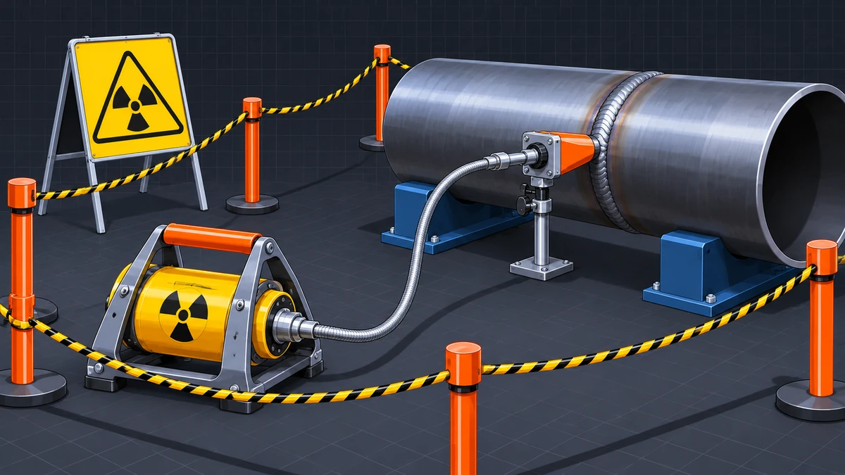
- Radiographers control dose with time, distance and shielding, and the dose rate from a gamma source falls with the square of the distance from it.
- Occupational dose limits are based on 20 mSv per year averaged over five years, with national differences in the single-year maximum, so always apply the limits of your regulator.
- Only certified radiographers working under a designated radiation safety officer should operate exposure devices, and assistants work under direct supervision.
- A calibrated survey meter must confirm that the source has returned to its shielded position after every exposure, before anyone approaches the device.
- Boundary distance depends on source activity, shielding and the limit used, so calculate it, confirm it by survey, and recalculate when the source changes.
- If a source cannot be retracted, or is damaged or lost, stop work, keep people away, never handle the source, and follow the written emergency procedure.
What Is Radiation Safety in Industrial Radiography and Why Does It Matter?
Radiation safety in industrial radiography is the system of equipment, procedures, monitoring and training that keeps the dose to workers and the public as low as reasonably achievable and below legal limits. It applies to gamma sources such as iridium-192 and to X-ray generators used for weld examination.
Radiation safety is a protection system that limits exposure to ionising radiation through justified use, optimised procedures and enforced dose limits.
Radiography is chosen because it shows volumetric flaws such as porosity and slag, which are described in the guide to welding defects, types, causes and remedies. The same penetrating radiation that reveals those flaws cannot be seen, felt or smelled, so people have no warning of a high dose rate unless they carry a working instrument.
Sealed gamma sources used in field radiography have high activity, so the dose rate near an unshielded source is very high. The IAEA safety guide on industrial radiography notes that the work poses negligible risk when done safely, but that workers and members of the public have been overexposed when sources were not controlled. Very high doses cause tissue reactions such as skin burns. Lower doses raise the probability of cancer later. Protection aims to prevent the first and minimise the second.
What Are the Three Principles of Radiation Protection?
The ICRP system rests on three principles: justification, optimisation and dose limitation. Justification means the practice does more good than harm, optimisation means doses are kept as low as reasonably achievable (ALARA), and dose limitation means individual doses stay below the limits set by the regulator.
What Radiation Sources Are Used in Industrial Radiography?
Industrial radiography uses either an X-ray generator or a sealed gamma source, most often iridium-192, selenium-75 or cobalt-60. The choice depends on material thickness, access and image quality, and it also decides the safety controls needed.
| Source | Radiation | Half-life | Typical steel thickness | Gamma constant, R m2/(Ci h) | Lead HVL |
|---|---|---|---|---|---|
| X-ray tube | X-rays, typically 100 to 400 kV | None; radiation only when energised | Thin to medium sections | Not applicable | Depends on kV |
| Selenium-75 | Gamma, about 0.1 to 0.4 MeV | About 120 days | About 3 to 29 mm | About 0.2 | About 2 mm |
| Iridium-192 | Gamma, about 0.2 to 0.6 MeV | About 74 days | About 12 to 63 mm | About 0.48 | About 5 mm |
| Cobalt-60 | Gamma, 1.17 and 1.33 MeV | About 5.3 years | About 50 to 150 mm | About 1.30 | About 12.5 mm |
Published values differ slightly between sources, so use the data sheet of your exposure device and the figures accepted by your regulator for any real calculation.
How Do X-ray and Gamma Sources Differ for Safety?
An X-ray generator produces radiation only while it is energised, so switching it off removes the hazard. A gamma source emits continuously and cannot be switched off, so it must stay in a shielded, locked exposure device between exposures. Gamma sources also decay, which changes the dose rate and boundary over time.
Many industrial radiography sources fall into Category 2 of the IAEA categorisation of radioactive sources, meaning they can be dangerous if not managed safely. Source inventories, locked storage, transport rules and prompt reporting of any loss are therefore core parts of radiography radiation safety.
What Are the Units and Dose Limits for Radiography Workers?
Dose limits cap the radiation dose a person may receive, expressed in millisieverts (mSv). The IAEA and ICRP framework sets 20 mSv per year averaged over five years for workers, with no more than 50 mSv in any single year, and 1 mSv per year for members of the public.
What Do Sievert, Gray and Roentgen Mean?
The gray (Gy) measures absorbed dose, and the sievert (Sv) measures dose weighted for radiation type and tissue. For X-rays and gamma rays, 1 Gy corresponds to about 1 Sv. Older instruments and texts use the roentgen (R) for exposure, and for gamma radiography it is practical to treat 1 R as about 10 mSv. Always check whether an instrument reads in mR/h, mSv/h or microsievert per hour before comparing a reading with a limit.
| Quantity | IAEA / ICRP | India (AERB) | USA (10 CFR Part 20) | EU Directive 2013/59/Euratom |
|---|---|---|---|---|
| Worker effective dose | 20 mSv/yr averaged over 5 years; max 50 mSv in any one year | 20 mSv/yr averaged over 5 years (sliding); 30 mSv in any year | 50 mSv (5 rem) per year | 20 mSv in any single year, with limited exceptions up to 50 mSv |
| Lens of the eye | 20 mSv/yr averaged over 5 years; max 50 mSv in any one year | 150 mSv/yr in the directive reviewed; a change to 20 mSv has been indicated | 150 mSv (15 rem) per year | 20 mSv in a year, or 100 mSv in 5 years with max 50 mSv in one year |
| Skin and extremities | 500 mSv per year | 500 mSv per year | 500 mSv (50 rem) per year | 500 mSv per year |
| Declared pregnancy, embryo or fetus | About 1 mSv for the rest of the pregnancy | 1 mSv for the rest of the pregnancy | 5 mSv (0.5 rem) for the pregnancy | About 1 mSv for the rest of the pregnancy |
| Member of the public | 1 mSv per year | 1 mSv per year | 1 mSv (0.1 rem) per year | 1 mSv per year |
Limits come from the IAEA basic safety standards and the ICRP recommendations, and each country adopts them in its own regulation. In India the Atomic Energy Regulatory Board (AERB) sets them, and in the United States the NRC and the Agreement States do. Apply the limits of the regulator that holds your licence, and check the current directive, because lens limits are being revised in several countries.
Limits are upper bounds, not targets. The ALARA principle requires employers to keep actual doses well below them.
How Do Time, Distance and Shielding Reduce Dose?
The three basic controls are time, distance and shielding: spend as little time as possible near the source, stay as far from it as practical, and place shielding between the source and people. Together with ALARA they reduce dose without relying on dose limits alone.
How Do Time and Distance Work?
Dose equals dose rate multiplied by time, so halving the time near a source halves the dose. For a small gamma source, dose rate falls with the square of distance, so doubling the distance cuts it to one quarter. This inverse square law makes a long crank-out cable and a large boundary the most effective site controls.
How Does Shielding Work?
Each half-value layer (HVL) of shielding material halves the dose rate. For iridium-192, approximate HVLs are about 5 mm of lead, about 13 mm of steel and about 43 to 45 mm of concrete. Three HVLs reduce the dose rate to one eighth, and ten HVLs to about one thousandth.
Exposure devices and collimators are forms of shielding. Pointing the beam toward the ground, a thick wall or an earth bank, and narrowing it with a collimator, can shrink the controlled area by a large factor.
How Is a Controlled Area Boundary Set for Site Radiography?
A controlled area boundary is the line, marked with ropes, signs and warning lights, outside which the dose rate stays below the limit used for unmonitored people. It is calculated before the job, confirmed with a calibrated survey meter during the first exposure, and watched throughout the work.
The boundary is rarely a circle, because shielding, walls and beam direction change the dose rate in each direction. Regulators commonly set the boundary limit to protect the public and define zones with higher dose rates that need access control. Table 6 later in this guide lists typical thresholds used in United States regulation. Check the thresholds in your own licence, and coordinate with other trades through the site permit system, as covered in the hot work permit system guide.
What Equipment and Monitoring Instruments Does a Radiographer Need?
Radiographers need a locked exposure device with guide tube and controls, a calibrated survey meter, an alarming ratemeter, direct-reading and passive dosimeters, and barriers with warning signs. Each item must be calibrated, tested or inspected before use, because a failed instrument gives false reassurance.
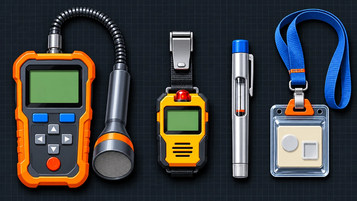
| Item | Purpose | Typical check or requirement |
|---|---|---|
| Exposure device (projector) | Holds the source in a shielded, locked position | Built to a recognised design standard, for example AERB/RF-IR/SS-1 (Rev.1) in India or ISO 3999; inspect lock, source indicator and condition before use |
| Guide tube, drive cable and collimator | Move the source to the exposure position and shape the beam | Inspect for damage, kinks and secure connections before each use |
| Survey meter | Measures dose rate and confirms source retraction | Calibrated at intervals not exceeding six months and after servicing under rules such as 10 CFR Part 34; function-checked before use |
| Alarming ratemeter | Sounds an alarm at a preset dose rate | Function check before each use; set the alarm level in the procedure |
| Direct-reading dosimeter | Shows accumulated dose during the job | Check zero and within calibration; read before and after each job |
| TLD, OSL or film badge | Provides the official dose record | Worn on the chest and exchanged each monitoring period through an approved service |
| Barriers, ropes, signs and lights | Mark and control the boundary | Visible from all approaches; lights or alarms where required |
| Source storage | Secure storage between jobs | Locked, labelled, with a log of every source movement |
In the United States, 10 CFR 34.47 requires a combination of a direct-reading dosimeter, an alarming ratemeter and a personnel dosimeter, and AERB practice requires a TLD badge worn at chest level. Lead aprons do not protect against the high-energy gamma rays of iridium-192 or cobalt-60. Distance, equipment shielding and a working survey meter are the real protection.
How Is a Site Radiography Job Controlled, Step by Step?
A site radiography job follows a fixed sequence: authorise, plan, set the boundary, check instruments, set up, expose, survey and record. Skipping the survey after retraction is what most often turns a routine job into an incident.
- Confirm authorisation and personnel. Check that the licence or permit covers the source, the site and the job, that only certified radiographers and trained assistants will work, and that the radiation safety officer knows about the job.
- Plan the exposure. Choose the source or X-ray unit, collimation, beam direction and timing, calculate the expected boundary distance, and arrange for other trades to be away from the area where possible.
- Establish the controlled area. Set barriers, ropes, signs and warning lights at the calculated boundary, confirm it by survey during the first exposure, and control every access point.
- Check instruments and monitoring. Confirm the survey meter and alarming ratemeter are in calibration and respond correctly, using a check source or reference reading at the start of each shift or as the regulator requires, and wear all required dosimeters.
- Set up the equipment. Position the exposure device, connect the guide tube and control cable, fit the collimator and place the film or detector, with the source locked in the shielded position.
- Sweep the area and expose. Make a final check that nobody is inside the boundary, give the agreed warning, wind out the source and keep the area under continuous observation.
- Retract and survey. Return the source to the shielded position, lock the device, and survey the device and guide tube with the survey meter to confirm the source is secure before anyone approaches.
- Record and secure. Record exposures, survey readings, dosimeter readings and any unusual event, then return the device to locked storage and update the source log.
Rules such as 10 CFR 34.41 require at least two qualified individuals at a temporary job site, and similar rules apply in many countries. Check what your regulator requires and never operate with an untrained helper.
Radiography fits into the inspection sequence defined in the inspection and test plan, and third-party inspectors often witness it, as described in the article on third party inspection in welding. Radiography inside a vessel or tank combines radiation and confined space hazards, so follow the confined space welding safety guide for entry, atmosphere testing and rescue arrangements as well as the radiation controls above.
Worked Example: How Far Should the Boundary Be for a 100 Ci Iridium-192 Source?
The boundary distance for an open, unshielded beam can be estimated with the inverse square law and the gamma constant. This example is for learning only, because real boundaries come from your radiation safety officer, licence conditions and survey readings.
What Are the Stated Inputs?
- Source: iridium-192, 100 Ci (3.7 TBq), half-life 73.8 days
- Gamma constant: 0.48 R m2/(Ci h), treated as about 4.8 mSv m2/(Ci h)
- Boundary limit: 0.02 mSv/h (2 mR/h), a common public-protection level
- Shield or collimator effect considered: 3 half-value layers
- Simplifications: point source, no scatter, build-up or air attenuation
Three half-value layers cut the boundary from about 155 m to about 55 m, and a source decayed to a quarter of its activity needs about half the distance. Boundaries must therefore be recalculated when the source changes. The conversion of 1 R to about 10 mSv is an approximation, and a real calculation uses the quantities and constants your regulator requires.
How Quickly Can Dose Accumulate?
Take the same unshielded 100 Ci source and a worker 20 m away in the open beam. The dose rate is 480 / 202 = 1.2 mSv/h, so a 5-minute exposure gives 1.2 x 5 / 60 = 0.1 mSv. At that rate, 200 exposures would reach 20 mSv. In practice workers stay outside the boundary and out of the beam, which is what the controls ensure.
What Should You Do in a Radiography Emergency?
If a source fails to return to its shielded position, is damaged, or cannot be accounted for, stop work, keep people away, never handle the source and follow the written emergency procedure. The priorities are to prevent exposure, control the area and call the radiation safety officer.
- Stop work and keep people back. Stop the exposure, move everyone away from the area and prevent anyone from entering the boundary.
- Do not touch the source or a damaged device. Do not pick up, move or disconnect any source or equipment unless the written emergency procedure and your training require it.
- Survey from a safe distance. Use the survey meter to find where the dose rate is high, mark the area and extend the boundary if readings exceed the limit.
- Keep the area under control. Post a person at each access point and keep signs and lights in place until the radiation safety officer clears the area.
- Notify. Inform the radiation safety officer at once and report to the regulator and the client as the licence and national rules require.
- Limit time near the source. Make only the entries needed to secure the source, using the shortest time, greatest distance and available shielding, and watch dosimeters.
- Record. Write down times, readings, positions, people present and their dosimeter results.
- Seek medical advice. Anyone who may have been overexposed, or who notices skin redness near a source, should be assessed by a doctor with the dosimetry results.
If anyone finds a metal capsule, device or container marked with the radiation trefoil, they should not touch or move it, should keep others away, and should report it at once to the site safety team, the regulator or the police so that a trained person can recover it.
Who Is Responsible for Radiation Safety and What Records Are Needed?
The organisation authorised to carry out radiography holds overall responsibility for radiation safety, and each role has defined duties. IAEA guidance names senior management, the radiation safety officer, radiographers and assistants, and it expects the roles to be written down.
| Role | Main responsibilities |
|---|---|
| Employer or licence holder | Holds the licence, appoints the radiation safety officer, and provides equipment, monitoring, training and procedures so that doses are kept as low as reasonably achievable |
| Radiation safety officer (RSO) | Ensures procedures are followed, reviews surveys and dose records, audits site work and leads the response to incidents |
| Certified radiographer | Operates the exposure device, sets and checks the boundary, performs surveys and supervises assistants |
| Radiographer’s assistant or intern | Works only under the direct supervision of a radiographer and does not operate the device alone |
| Client or site owner | Provides access, coordinates other trades and permits, and supports the boundary |
| Other site workers | Respect signs and boundaries, report breaches and never enter a controlled area |
Radiographers are trained and certified through national schemes, and the article on NDT Level 1, 2 and 3 certification explains how NDT personnel qualification differs from radiation safety certification. QA/QC engineers and inspectors who witness radiography, covered in the guide to welding engineer, QA/QC engineer and welding inspector roles, must follow the same boundary rules as the radiography team.
Which Records Should Be Kept?
- Personal dose records from the monitoring service, and direct-reading dosimeter logs for each job.
- Calibration and function-check records for survey meters and alarming ratemeters.
- Source inventory, movement log, leak test and device inspection records.
- Training, certification and refresher records for each radiographer and assistant.
- Job records showing source, boundary, survey readings and any unusual event.
- Incident and investigation reports, with corrective actions.
These records belong in the same controlled system as weld maps and test reports, as described in the guide to welding documentation and record-keeping.
How Is Industrial Radiography Regulated in India and Elsewhere?
Every country regulates radiography through a national authority, and most base their rules on the IAEA basic safety standards. In India that authority is the Atomic Energy Regulatory Board (AERB), which licenses sources, approves radiation safety officers and certifies radiographers.
| Jurisdiction | Regulator | Key documents | Notable requirements |
|---|---|---|---|
| India | Atomic Energy Regulatory Board (AERB) | Atomic Energy (Radiation Protection) Rules, 2004; AERB/RF-IR/SC-1 (Rev.1) Industrial Radiography (2016, amended 2019); AERB/RF-IR/SS-1 (Rev.1) exposure devices and source changers | Licence for sources, AERB-approved RSO, certified radiographers, personnel monitoring with TLD |
| United States | NRC and Agreement States | 10 CFR Part 34 and 10 CFR Part 20 | Radiographer certification, RSO, two qualified persons at temporary sites, survey meter calibration, personnel monitoring |
| European Union | National authorities | Directive 2013/59/Euratom, applied in national law | Dose limits, authorisation of practices, qualified experts and training |
| United Kingdom | Health and Safety Executive | Ionising Radiations Regulations 2017 | Authorisation of work, radiation protection adviser, dose limits and local rules |
| United Arab Emirates | Federal Authority for Nuclear Regulation (FANR) | FANR regulations; confirm the current list | Licensing of sources and facilities, trained personnel |
| International | IAEA | GSR Part 3; SSG-11 | Basis for national rules; SSG-11 covers duties, safety assessment, training, monitoring, site radiography, transport and emergencies |
Contracts often add requirements for notification, supervision and reporting. Confirm the current regulation, licence conditions and contract terms before work begins.
Quick Reference: Dose-Rate Thresholds and Pre-Exposure Checklist
Table 6 lists dose-rate thresholds used in United States regulation, as an example of how zones are defined. Other regulators use different wording and values, so treat the table as a guide to the concept, not as a limit for your site.
| Zone or level | Typical definition | Usual action |
|---|---|---|
| Public boundary | Not more than 0.02 mSv (2 mrem) in any one hour in unrestricted areas | Keep unmonitored people outside this boundary |
| Radiation area | More than 0.05 mSv/h (5 mrem/h) at 30 cm from the source | Post warning signs and restrict access |
| High radiation area | More than 1 mSv/h (100 mrem/h) at 30 cm from the source | Control entry with barriers, locks or alarms |
| Very high radiation area | 5 Gy/h (500 rad/h) or more at 1 m from the source | Apply additional access controls because severe injury can occur quickly |
- Licence, source and personnel authorised for this job and site.
- Boundary calculated, marked and visible from every approach.
- Survey meter and alarming ratemeter in calibration and working.
- Direct-reading dosimeter zeroed and TLD badge worn.
- Exposure device, guide tube and collimator inspected and connected.
- Other trades informed and nobody inside the boundary.
- Emergency procedure and contact numbers at hand.
What Are the Common Mistakes in Radiography Radiation Safety?
Most incidents follow a few repeated procedural errors, and each is preventable with the controls described above.
- Not surveying the device and guide tube after the source is retracted.
- Letting an untrained assistant operate the device.
- Setting the boundary from a rule of thumb instead of a calculation and survey.
- Forgetting that a new or decayed source changes the boundary distance.
- Working without an alarming ratemeter or dosimeter, or with an out-of-calibration survey meter.
- Using a damaged guide tube, drive cable or locking mechanism.
- Leaving an exposure area unattended or poorly signed.
- Not telling other trades when exposures will take place.
- Storing a source without a lock or movement log.
- Trying to free a jammed source by hand instead of following the emergency procedure.
Requirements depend on the regulation, licence conditions and contract specification applicable to your site.
Key Terms
- Sievert (Sv)
- The unit of effective or equivalent dose that accounts for the type of radiation and the tissue exposed.
- ALARA
- The principle that doses are kept as low as reasonably achievable, with economic and social factors taken into account.
- Controlled area
- An area where access is restricted and special protection measures apply during radiography.
- Half-value layer (HVL)
- The thickness of a material that reduces the dose rate to one half.
- Survey meter
- A calibrated portable instrument that measures dose rate and confirms the source position.
- Alarming ratemeter
- A personal instrument that sounds an alarm when the dose rate exceeds a preset level.
- Exposure device
- A shielded, locked container that holds a sealed gamma source when it is not exposed.
- Radiation safety officer (RSO)
- The designated person responsible for radiation safety procedures, monitoring and incident response.
Frequently Asked Questions About Radiography Radiation Safety
What is the annual dose limit for a radiographer?
Limits depend on the regulator. The IAEA and ICRP framework is 20 mSv per year averaged over five years, with no more than 50 mSv in a single year. In India, AERB applies 20 mSv per year averaged over five years and 30 mSv in any year, and the United States uses 50 mSv per year. Doses should be kept well below the limits, and the dose record must show each worker’s total.
How far should the boundary be for gamma radiography?
There is no fixed distance. It depends on source activity, collimation, shielding, beam direction and the limit used at the boundary. For an open, unshielded 100 Ci iridium-192 source, a simple inverse square estimate gives about 155 m for 0.02 mSv/h. The real boundary must be confirmed with a calibrated survey meter during the first exposure.
Is X-ray radiography safer than gamma radiography?
An X-ray generator stops emitting when it is switched off, which removes the risk of an unretracted source and the need to store a radioactive source. It still needs a controlled boundary, warning signals and trained operators. Gamma sources need locked storage, accounting and transport controls. Neither method is safe without correct procedures, and the choice also depends on thickness and access.
Why must the source and device be surveyed after every exposure?
The survey meter reading is the most reliable proof that the source has returned to its shielded position. Indicators on the device can be wrong if the source or cable is damaged. Approaching a device without a survey can lead to accidental overexposure. The survey should cover the device and the guide tube before anyone enters the boundary.
Can a pregnant worker do radiography?
Dose limits for a declared pregnancy are lower, about 1 mSv for the remainder of the pregnancy under the IAEA, ICRP and AERB approach and 5 mSv under United States rules. Employers should discuss duties with the worker once a pregnancy is declared and adjust work so that the limit cannot be exceeded. Rules vary, so follow the national regulation and the advice of the radiation safety officer.
Do lead aprons protect radiographers?
Not against the high-energy gamma rays of iridium-192 or cobalt-60. These rays pass through thin lead, so an apron gives little protection and can create false confidence. Radiographers rely on distance, equipment shielding, collimation, procedures and personal monitoring. Ask the radiation safety officer for guidance on any special case.
Conclusion
Radiography radiation safety depends on layered controls: a licensed source, certified people, a calculated and surveyed boundary, calibrated instruments and a survey after every exposure. The worked example shows why distance and shielding matter, because three half-value layers cut the boundary for a 100 Ci iridium-192 source from about 155 m to about 55 m. Keep doses well below the limits, record every exposure, and rehearse the emergency procedure before it is needed, so that nobody has to improvise during an incident. As a next step, test your knowledge of radiographic technique and safety with the radiography quiz, and review your licence conditions with your radiation safety officer.
Standards and References
- IAEA Safety Standards Series No. GSR Part 3, Radiation Protection and Safety of Radiation Sources: International Basic Safety Standards (IAEA)
- IAEA Safety Standards Series No. SSG-11, Radiation Safety in Industrial Radiography (IAEA, 2011; a revision is in preparation, so check the current edition)
- IAEA Safety Standards Series No. RS-G-1.9, Categorization of Radioactive Sources (IAEA)
- ICRP Publication 103, The 2007 Recommendations of the International Commission on Radiological Protection, and ICRP Publication 118, Statement on Tissue Reactions (ICRP)
- Atomic Energy (Radiation Protection) Rules, 2004, and AERB Safety Directive on dose limits for workers and the public (2011) (Government of India, AERB)
- AERB/RF-IR/SC-1 (Rev.1), Safety Code on Industrial Radiography (AERB, 2016, amended 2019)
- AERB/RF-IR/SS-1 (Rev.1), Industrial Gamma Radiography Exposure Devices and Source Changers (AERB)
- 10 CFR Part 20, Standards for Protection Against Radiation, and 10 CFR Part 34, Licenses for Industrial Radiography and Radiation Safety Requirements for Industrial Radiographic Operations (US NRC)
- Directive 2013/59/Euratom, basic safety standards for protection against ionising radiation (European Union)
- ISO 3999, Radiation protection – Apparatus for industrial gamma radiography – Specifications for performance, design and tests (ISO)
- ISO 17636-1 and ISO 17636-2, Non-destructive testing of welds – Radiographic testing (ISO)
Requirements depend on the regulation, licence conditions and contract specification applicable to your site.
This guide was prepared by the WeldFabWorld technical team from the standards and regulator publications listed under Standards and References. It is general information and does not replace licence conditions or the instructions of your radiation safety officer.
Related Guides on WeldFabWorld
More guides on workplace safety are collected in the Safety & Compliance category.